von Ana Catarina Ribeiro Carrão
Statistik und Sichtungsnachweis dieser Seite findet sich am Artikelende
| [1.] Arc/Fragment 041 01 - Diskussion Zuletzt bearbeitet: 2014-02-26 23:07:05 Guckar | Arc, Fragment, Gesichtet, SMWFragment, Schutzlevel sysop, Toyota et al 2005, Verschleierung |
|
|
|
| Untersuchte Arbeit: Seite: 41, Zeilen: 1-22 |
Quelle: Toyota et al 2005 Seite(n): 2109, Zeilen: l.col: 46ff |
|---|---|
| [After the surgery, analgesic] (buprenorphine 0.05 mg/kg s.c.) and antibiotic (enrofloxacin 10 mg/kg s.c.) compounds were administered. Rats were observed in a recovery cage for 2 hours and then transferred to the animal care facility. For 3 days after the surgery, buprenorphine (0.5 mg/kg BID mixed in strawberry Jello) was taken orally for pain relief. On the fourth day after the surgery, the ischemic protocol was started (see the Experimental Protocol section). After 5 days of the experimental protocol, the rats were anesthetized and the chest was opened by mid thoracotomy. In CBF measurement or DHE analysis groups, the hearts were excised at the end of procedures, and the tissue was prepared for analyses.
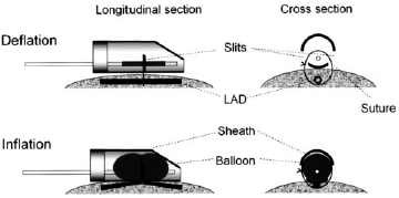
3.2 Mini-Pneumatic Snare Occluder for Rat Heart A mini-pneumatic snare occluder consisting of a mini-balloon, sheath tubing, suture, and catheter (Fig. 11) was placed on the LAD of the rat heart. The balloon (7 mm long) is made of soft latex membrane and is sufficiently pliable to give negligible physical force on the coronary vessels during balloon deflation. The balloon is mounted within an umbrella sheath (3.2 or 4.8 mm in diameter, 12 mm in length; protects the balloon from fibrous infiltration). Prolene (5– 0) is passed around the LAD and attached to the sheath, securing the occluder to the heart, so that myocardial ischemia is produced by balloon inflation. Inflation volume is small (0.2 to 0.25 mL air), but occlusion occurs by 2 physical actions: “crimping” the LAD toward upward/outside and compressing the LAD by the inflated balloon/sheath. The balloon is connected to a catheter (PE-50) that is exteriorized. Balloon inflation and deflation are controlled from outside the rat cage.
Fig. 11 - Schematic diagram of the mini-pneumatic snare and its actions. Top: Cross-sectional and longitudinal views when the balloon is deflated. Bottom: Views during inflation. The artery is patent when the balloon is deflated, but during inflation, a snare situated underneath the artery is pulled “upward” during inflation, producing the coronary occlusion94. 94. Kappel A, Ronicke V, Damert A, Flamme I, Risau W, Breier G. Identification of Vascular Endothelial Growth Factor (VEGF) Receptor-2 (Flk-1) Promoter/Enhancer Sequences Sufficient for Angioblast and Endothelial Cell- Specific Transcription in Transgenic Mice. Blood. 1999;93:4284-4292. |
After the surgery, analgesic (buprenorphine 0.05 mg/kg SC) and antibiotic (enrofloxacin 10 mg/kg SC) were administered. Rats were observed in a recovery cage for 2 hours and then transferred to the animal care facility. For 3 days after the surgery, buprenorphine (0.5 mg/kg BID mixed in strawberry Jello) was taken orally for pain. On the fourth day after the surgery, ischemic protocol was started (see the Experimental Protocol section).
After 10 days of the experimental protocol, the rats were anesthetized, and the chest was opened by mid thoracotomy. In the micro-CT group, the hearts were immediately excised. In CBF measurement group, blood flow to the normal and collateraldependent zones during coronary occlusion was measured. The heart was excised at the end of measurements, and the tissue was prepared for analyses of radioactivity. Mini-Pneumatic Snare Occluder for Rat Heart We developed a mini-pneumatic snare occluder (patent application serial number: 11/071,617, E.T. and W.M.C.) consisting of a mini-balloon, sheath tubing, suture, and catheter (Figure 1). The balloon (7 mm long) is made of soft latex membrane and is sufficiently pliable to give negligible physical force on the coronary vessels during balloon deflation. The balloon is mounted within an umbrella sheath (3.2 or 4.8 mm in diameter, 12 mm in length; protects the balloon from fibrous infiltration). Prolene (5– 0) is passed around the LAD and attached to the sheath, securing the occluder to the heart, so that myocardial ischemia is produced by balloon inflation. Inflation volume is small (0.2 to 0.25 mL air), but occlusion occurs by 2 physical actions: “crimping” the LAD toward upward/outside and compressing the LAD by the inflated balloon/sheath. The balloon is connected to a catheter (PE-50) that is exteriorized. Balloon inflation and deflation are controlled from outside the rat cage.
Figure 1. Schematic of the mini-pneumatic snare and its actions. Top, Cross-sectional and longitudinal views when the balloon is deflated. Bottom, Views during inflation. The artery is patent when the balloon is deflated, but during inflation, a snare situated underneath the artery is pulled “upward” during inflation, producing the coronary occlusion. |

At the beginning of the previous page the source is mentioned, but without indication that the following two pages are taken from it. The given source Kappel et al. (1999) doesn't contain any of the copied material. |
|
Letzte Bearbeitung dieser Seite: durch Benutzer:Hindemith, Zeitstempel: 20140225220156