|
|
|
| Untersuchte Arbeit: Seite: 21, Zeilen: 1ff (komplett) |
Quelle: Moor instruments 2006 Seite(n): 2, Zeilen: 5ff |
|---|---|
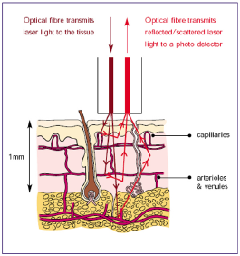
| Normal fibre separations in the probe head are a few tenths of 1mm, consequently blood flow is measured in a tissue volume of typically 1mm3 or smaller.
(Fig.1.8.a)
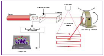
(Basic Theory and Operating Principles of LDF & LDI, Moor instruments Ltd User manual, 2003) In a Laser Doppler blood flow Imager (LDI) the low intensity laser beam is scanned across a tissue surface in a raster fashion using a moving mirror. There is no direct contact with the tissue being assessed. The basic elements of the moorLDI are shown schematically in the following figure. (Fig.1.8.b)
(Basic Theory and Operating Principles of LDF & LDI, Moor instruments Ltd User manual, 2003) |
Normal fibre separations in the probe head are a few tenths of a mm, consequently blood flow is measured in a tissue volume of typically 1mm3 or smaller. [...]
(c)
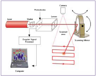
In a Laser Doppler blood flow Imager (LDI) the low intensity laser beam is scanned across a tissue surface in a raster fashion using a moving mirror. There is no direct contact with the tissue being assessed. The basic elements of the moorLDI are shown schematically in the following figure. (d)
|

Für die Abbildungen ist die Quelle angegeben, für den Text nicht. |
|